Biological Structures
BIOLOGICAL SAMPLES
For many years biologists have looked at structures in organisms and their cells with polarized light microscopy. The birefringence that occurs inside these organized biological structures offers a way to see them without staining or dying, and some structures are only visible because of their birefringence. Polarized light microscopy offered a valuable if qualitative tool to identify things like spindle fibers and myelination.
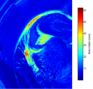
With the advent of cameras for imaging microscopy has come an ability to image and quantitate birefringence in structures. From muscle fibers in mosquito larvae to myelination in mouse brain tissue, Hinds’ Exicor® MicroImagerTM offers not only a fine image of birefringence structures, but also the ability to quantitate both the birefringence and the angle of the fast axis that it creates.
SUGGESTED PRODUCT
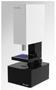
Exicor® MicroImagerTM
Exicor® MicroImagerTM Product Bulletin
Figure 1 (left). Myelinated White Matter in a mouse brain.
Figure 2 (right). Birefringence imaged using the Exicor® MicroImagerTM.
Contact us to see how Hinds Instruments works with our customers to solve complex metrology problems.

